Home
/ Posterior Neck Muscle Diagram, Anterior Triangle of the Neck at University Of British ..., The neck muscles, including the sternocleidomastoid and the trapezius, are responsible for the gross motor movement in the muscular system of the head and neck.
Posterior Neck Muscle Diagram, Anterior Triangle of the Neck at University Of British ..., The neck muscles, including the sternocleidomastoid and the trapezius, are responsible for the gross motor movement in the muscular system of the head and neck.
Posterior Neck Muscle Diagram, Anterior Triangle of the Neck at University Of British ..., The neck muscles, including the sternocleidomastoid and the trapezius, are responsible for the gross motor movement in the muscular system of the head and neck.. Outer surface of second rib action scalenus posterior muscle. The muscle consists of three parts which fan out during their course This site contains information about posterior neck muscle anatomy. Start studying posterior neck muscles. The muscles in the posterior compartment of the forearm are commonly known as the extensor muscles.
You've got anterior, middle and posterior scalene muscles. This site contains information about posterior neck muscle anatomy. It's got a superior, inferior, oblique part and a vertical part. Bounding a large anatomic region the posterior neck triangle further divides into two smaller triangles by the inferior omohyoid muscle. It is the smallest of the scalene muscles and situated most profoundly.
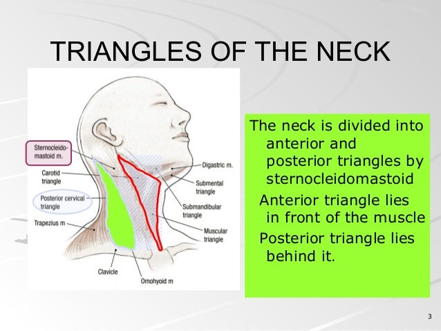
Medicowesome: Triangles of the neck diagram and mnemonic from lh6.ggpht.com The muscles of the neck can be divided into posterior and anterior groups: This muscle diagram is interactive: The posterior triangle has the following boundaries: They are all innervated by the radial nerve. Outer surface of second rib action scalenus posterior muscle. In the posterior triangle the spinal accessory nerve is adherent to the deep aspect of the fascial roof (formed by prevertebral layer of deep cervical fascia) of the triangle and is surrounded by lymph nodes. The muscles in the posterior compartment of the forearm are commonly known as the extensor muscles. (supplies deep muscles of the neck).
The posterior triangle has the following boundaries:
This chart includes views of the posterior thoracic wall in five separate illustrations, and almost a dozen views of back of the neck/head, including three closeup views. The neck muscles, including the sternocleidomastoid and the trapezius, are responsible for the gross motor movement in the muscular system of the head and neck. Rectus capitis posterior major, rectus capitis posterior minor, obliquus capitis superior, obliquus capitis inferior. It is deeply placed, lying behind sternocleidomastoid. Click here for a diagram of the the posterior belly of digastric muscle and its relations. Start studying posterior neck muscles. Posterior border of the sternocleidomastoideus. The next muscle is the splenius capitis. Advertisements help pay for this website. Quickly memorize the terms, phrases and much more. Trapezius is a large, paired, triangular shaped muscle located in the upper back and neck. The masseter muscle originates on the zygomatic arch, and inserts onto. Muscles, connected to bones or internal but muscle is also the dominant tissue in the heart and in the walls of other hollow organs of the body.
It is the smallest of the scalene muscles and situated most profoundly. Rectus capitis posterior major, rectus capitis posterior minor, obliquus capitis superior, obliquus capitis inferior. You've got anterior, middle and posterior scalene muscles. This muscle diagram is interactive: Working in pairs on the left and.
posterior triangle of neck from image.slidesharecdn.com Thank you for your support. Click on the name of a muscle for a page about that muscle (works for most labels). Bounding a large anatomic region the posterior neck triangle further divides into two smaller triangles by the inferior omohyoid muscle. The neck muscles, including the sternocleidomastoid and the trapezius, are responsible for the gross motor movement in the muscular system of the head and neck. I'll just flick over to a diagram to show you these, but they originate on the transverse processes of what this muscle does is that it flexes the neck anteriorly. Whiplash associated disorders and neck rehabilitation review whiplash and the related management of the cervical spine. Muscles, connected to bones or internal but muscle is also the dominant tissue in the heart and in the walls of other hollow organs of the body. In addition, the posterior neck muscles may be damaged during the hyperflexion phase.
Their main function is contractibility.
Advertisements help pay for this website. Your posterior neck muscles are those muscles that lie within the posterior triangle of the neck, beneath that investing layer of fascia, although they are not the only. It's got a superior, inferior, oblique part and a vertical part. Thank you for your support. Learn vocabulary, terms and more with flashcards, games and other study tools. In the posterior triangle the spinal accessory nerve is adherent to the deep aspect of the fascial roof (formed by prevertebral layer of deep cervical fascia) of the triangle and is surrounded by lymph nodes. The posterior scalene is the smallest and deepest of the scalene muscles. These muscles form a small slip on each side, which is nearly parallel to the posterior belly of the digastric muscle. I have also done a tutorial on the anterior triangle of the neck, so please watch that if you are interested! This tutorial covers the muscles of the posterior triangle of the neck as well as the prevertebral and lateral neck muscles. Posterior muscles in the body. Assoc prof craig hacking ◉ ◈ and dr vijay et al. The next muscle is the splenius capitis.
Early & selective in disease: Posterior muscles in the body. Anteriorly by posterior border of sternocleidomastoid muscle. Their main function is contractibility. Thank you for your support.
muscles of head and neck - Anatomy & Physiology 2200 with ... from s3.amazonaws.com Click here for a diagram of the the posterior belly of digastric muscle and its relations. Posterior border of the sternocleidomastoideus. Trapezius is a large, paired, triangular shaped muscle located in the upper back and neck. Thank you for your support. They are all innervated by the radial nerve. The posterior triangle has the following boundaries: (supplies deep muscles of the neck). Although this division is not perfect (e.g., the similarly, all muscles that cross the spinal joints posteriorly are extensors of the neck at the spinal joints.
Thank you for your support.
Posterior compartment of the leg leg muscles anterior posterior lateral view ankleanatomymuscleap gif ankleanatomymuscleposterior gif. They move the head in every direction, pulling the skull and jaw towards the shoulders, spine, and scapula. This muscle diagram is interactive: Thank you for your support. The posterior triangle is bounded: Learn vocabulary, terms and more with flashcards, games and other study tools. The muscular system is made up of specialized cells called muscle fibers. Start studying posterior neck muscles. Whiplash associated disorders and neck rehabilitation online course: This tutorial covers the muscles of the posterior triangle of the neck as well as the prevertebral and lateral neck muscles. The muscle consists of three parts which fan out during their course Advertisements help pay for this website. This muscle has three parts.
Fortunately, these muscles, including the posterior neck muscles, can be described in ways that are fairly easy to understand neck muscle diagram. (supplies deep muscles of the neck).

